Bones & Born Again Diane Cluck
| Bone | |
|---|---|
 A bone dating from the Pleistocene Ice Age of an extinct species of elephant | |
 A scanning electronic micrograph of bone at x,000× magnification | |
| Identifiers | |
| MeSH | D001842 |
| TA98 | A02.0.00.000 |
| TA2 | 366, 377 |
| TH | H3.01.00.0.00001 |
| FMA | 5018 |
| Anatomical terminology [edit on Wikidata] | |
A bone is a rigid organ[ane] that constitutes part of the skeleton in nearly vertebrate animals. Bones protect the various other organs of the trunk, produce red and white blood cells, store minerals, provide structure and support for the body, and enable mobility. Bones come up in a variety of shapes and sizes and accept a circuitous internal and external construction.[ii] They are lightweight yet potent and hard, and serve multiple functions.
Bone tissue (osseous tissue) is difficult tissue, a blazon of specialized connective tissue. It has a honeycomb-similar matrix internally, which helps to requite the os rigidity. Bone tissue is made upwards of dissimilar types of bone cells. Osteoblasts and osteocytes are involved in the formation and mineralization of bone; osteoclasts are involved in the resorption of os tissue. Modified (flattened) osteoblasts become the lining cells that grade a protective layer on the os surface. The mineralized matrix of os tissue has an organic component of mainly collagen called ossein and an inorganic component of bone mineral made upwardly of various salts. Bone tissue is mineralized tissue of two types, cortical os and cancellous bone. Other types of tissue constitute in basic include bone marrow, endosteum, periosteum, fretfulness, blood vessels and cartilage.
In the human torso at nascency, there are approximately 300 bones present; many of these fuse together during development, leaving a total of 206 separate bones in the adult, not counting numerous small sesamoid basic.[iii] [iv] [5] The largest bone in the torso is the femur or thigh-bone, and the smallest is the stapes in the middle ear.
The Greek word for os is ὀστέον ("osteon"), hence the many terms that use it as a prefix—such as osteopathy.
Structure [edit]
Os is not uniformly solid, but consists of a flexible matrix (near 30%) and leap minerals (about 70%) which are intricately woven and incessantly remodeled by a grouping of specialized bone cells. Their unique composition and design allows bones to be relatively hard and strong, while remaining lightweight.
Os matrix is 90 to 95% composed of rubberband collagen fibers, also known as ossein,[six] and the remainder is ground substance.[7] The elasticity of collagen improves fracture resistance.[viii] The matrix is hardened past the binding of inorganic mineral table salt, calcium phosphate, in a chemical arrangement known as calcium hydroxylapatite. Information technology is the bone mineralization that requite bones rigidity.
Bone is actively constructed and remodeled throughout life by special bone cells known every bit osteoblasts and osteoclasts. Inside whatever single os, the tissue is woven into 2 main patterns, known equally cortical and cancellous os, and each with dissimilar appearance and characteristics.
Cortex [edit]

Cross-section details of a long os
The difficult outer layer of bones is composed of cortical os, which is also called compact bone equally it is much denser than cancellous bone. It forms the hard exterior (cortex) of bones. The cortical bone gives bone its smooth, white, and solid advent, and accounts for fourscore% of the total bone mass of an adult homo skeleton.[9] Information technology facilitates bone'due south principal functions—to back up the whole body, to protect organs, to provide levers for motility, and to store and release chemical elements, mainly calcium. It consists of multiple microscopic columns, each called an osteon or Haversian system. Each column is multiple layers of osteoblasts and osteocytes effectually a cardinal canal called the haversian canal. Volkmann'due south canals at right angles connect the osteons together. The columns are metabolically active, and as bone is reabsorbed and created the nature and location of the cells within the osteon volition change. Cortical os is covered by a periosteum on its outer surface, and an endosteum on its inner surface. The endosteum is the purlieus between the cortical bone and the cancellous bone.[10] The main anatomical and functional unit of cortical bone is the osteon.
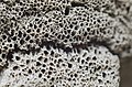
Trabecules [edit]
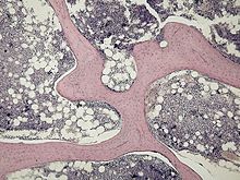
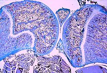
Micrograph of cancellous bone
Cancellous os, also called trabecular or spongy bone,[ten] is the internal tissue of the skeletal os and is an open up prison cell porous network. Cancellous bone has a higher surface-area-to-volume ratio than cortical bone and it is less dense. This makes it weaker and more flexible. The greater surface surface area also makes it suitable for metabolic activities such equally the exchange of calcium ions. Cancellous bone is typically constitute at the ends of long bones, near joints and in the interior of vertebrae. Cancellous bone is highly vascular and ofttimes contains scarlet bone marrow where hematopoiesis, the product of claret cells, occurs. The primary anatomical and functional unit of cancellous os is the trabecula. The trabeculae are aligned towards the mechanical load distribution that a bone experiences within long bones such as the femur. As far as short bones are concerned, trabecular alignment has been studied in the vertebral pedicle.[11] Thin formations of osteoblasts covered in endosteum create an irregular network of spaces,[12] known as trabeculae. Within these spaces are bone marrow and hematopoietic stem cells that requite rise to platelets, cherry claret cells and white blood cells.[12] Trabecular marrow is composed of a network of rod- and plate-like elements that brand the overall organ lighter and permit room for claret vessels and marrow. Trabecular bone accounts for the remaining 20% of full bone mass but has nearly ten times the surface area of compact bone.[13]
The words cancellous and trabecular refer to the tiny lattice-shaped units (trabeculae) that form the tissue. Information technology was first illustrated accurately in the engravings of Crisóstomo Martinez.[14]
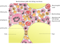
Marrow [edit]
Bone marrow, also known equally myeloid tissue in red bone marrow, can be found in almost any bone that holds cancellous tissue. In newborns, all such bones are filled exclusively with cherry-red marrow or hematopoietic marrow, merely equally the child ages the hematopoietic fraction decreases in quantity and the fatty/ yellow fraction called marrow adipose tissue (MAT) increases in quantity. In adults, red marrow is mostly found in the bone marrow of the femur, the ribs, the vertebrae and pelvic basic.[15]
Cells [edit]

Bone is metabolically active tissue composed of several types of cells. These cells include osteoblasts, which are involved in the creation and mineralization of os tissue, osteocytes, and osteoclasts, which are involved in the reabsorption of bone tissue. Osteoblasts and osteocytes are derived from osteoprogenitor cells, but osteoclasts are derived from the same cells that differentiate to form macrophages and monocytes.[16] Within the marrow of the bone there are also hematopoietic stalk cells. These cells give rise to other cells, including white blood cells, red blood cells, and platelets.[17]
Osteoblast [edit]

Light micrograph of decalcified cancellous bone tissue displaying osteoblasts actively synthesizing osteoid, containing two osteocytes.
Osteoblasts are mononucleate bone-forming cells. They are located on the surface of osteon seams and make a poly peptide mixture known as osteoid, which mineralizes to become bone.[18] The osteoid seam is a narrow region of newly formed organic matrix, not still mineralized, located on the surface of a bone. Osteoid is primarily composed of Type I collagen. Osteoblasts too manufacture hormones, such as prostaglandins, to act on the bone itself. The osteoblast creates and repairs new bone past actually building effectually itself. Starting time, the osteoblast puts upwards collagen fibers. These collagen fibers are used as a framework for the osteoblasts' piece of work. The osteoblast then deposits calcium phosphate which is hardened by hydroxide and bicarbonate ions. The make-new os created past the osteoblast is called osteoid.[xix] Once the osteoblast is finished working it is actually trapped inside the os one time it hardens. When the osteoblast becomes trapped, it becomes known as an osteocyte. Other osteoblasts remain on the top of the new os and are used to protect the underlying bone, these go known equally lining cells.[ citation needed ]
Osteocyte [edit]
Osteocytes are cells of mesenchymal origin and originate from osteoblasts that take migrated into and get trapped and surrounded by bone matrix that they themselves produced.[10] The spaces the cell body of osteocytes occupy within the mineralized collagen blazon I matrix are known as lacunae, while the osteocyte cell processes occupy channels called canaliculi. The many processes of osteocytes reach out to meet osteoblasts, osteoclasts, bone lining cells, and other osteocytes probably for the purposes of communication.[20] Osteocytes remain in contact with other osteocytes in the bone through gap junctions—coupled jail cell processes which pass through the canalicular channels.
Osteoclast [edit]
Osteoclasts are very large multinucleate cells that are responsible for the breakdown of bones by the procedure of bone resorption. New bone is then formed by the osteoblasts. Bone is constantly remodeled by the resorption of osteoclasts and created by osteoblasts.[16] Osteoclasts are large cells with multiple nuclei located on bone surfaces in what are called Howship's lacunae (or resorption pits). These lacunae are the event of surrounding bone tissue that has been reabsorbed.[21] Considering the osteoclasts are derived from a monocyte stem-cell lineage, they are equipped with phagocytic-similar mechanisms similar to circulating macrophages.[16] Osteoclasts mature and/or migrate to discrete bone surfaces. Upon arrival, active enzymes, such as tartrate-resistant acid phosphatase, are secreted confronting the mineral substrate.[ commendation needed ] The reabsorption of bone past osteoclasts also plays a role in calcium homeostasis.[21]
Composition [edit]
Basic consist of living cells (osteoblasts and osteocytes) embedded in a mineralized organic matrix. The chief inorganic component of human bone is hydroxyapatite, the dominant bone mineral, having the nominal composition of Ca10(PO4)6(OH)2.[22] The organic components of this matrix consist mainly of type I collagen—"organic" referring to materials produced as a result of the human body—and inorganic components, which alongside the dominant hydroxyapatite phase, include other compounds of calcium and phosphate including salts. Approximately 30% of the acellular component of bone consists of organic affair, while roughly lxx% by mass is attributed to the inorganic phase.[23] The collagen fibers give os its tensile strength, and the interspersed crystals of hydroxyapatite give bone its compressive forcefulness. These furnishings are synergistic.[23] The exact limerick of the matrix may be subject to change over time due to nutrition and biomineralization, with the ratio of calcium to phosphate varying between 1.three and 2.0 (per weight), and trace minerals such as magnesium, sodium, potassium and carbonate also being establish.[23]
Type I collagen composes xc–95% of the organic matrix, with remainder of the matrix being a homogenous liquid called ground substance consisting of proteoglycans such every bit hyaluronic acrid and chondroitin sulfate,[23] equally well as non-collagenous proteins such as osteocalcin, osteopontin or bone sialoprotein. Collagen consists of strands of repeating units, which give bone tensile strength, and are arranged in an overlapping fashion that prevents shear stress. The role of ground substance is not fully known.[23] Ii types of os tin be identified microscopically according to the arrangement of collagen: woven and lamellar.
- Woven bone (too known as gristly os), which is characterized by a haphazard organization of collagen fibers and is mechanically weak.[24]
- Lamellar bone, which has a regular parallel alignment of collagen into sheets ("lamellae") and is mechanically stiff.[24]
Woven bone is produced when osteoblasts produce osteoid rapidly, which occurs initially in all fetal basic, but is afterward replaced by more resilient lamellar bone. In adults woven bone is created after fractures or in Paget'south affliction. Woven bone is weaker, with a smaller number of randomly oriented collagen fibers, only forms quickly; it is for this advent of the fibrous matrix that the bone is termed woven. Information technology is before long replaced by lamellar os, which is highly organized in concentric sheets with a much lower proportion of osteocytes to surrounding tissue. Lamellar bone, which makes its first appearance in humans in the fetus during the third trimester,[25] is stronger and filled with many collagen fibers parallel to other fibers in the same layer (these parallel columns are called osteons). In cross-section, the fibers run in opposite directions in alternate layers, much like in plywood, profitable in the bone's ability to resist torsion forces. After a fracture, woven bone forms initially and is gradually replaced by lamellar bone during a process known as "bony commutation." Compared to woven bone, lamellar os formation takes place more slowly. The orderly deposition of collagen fibers restricts the formation of osteoid to nearly 1 to 2 µm per twenty-four hour period. Lamellar bone besides requires a relatively apartment surface to lay the collagen fibers in parallel or concentric layers.[26]
Deposition [edit]
The extracellular matrix of os is laid down past osteoblasts, which secrete both collagen and ground substance. These synthesise collagen within the prison cell and and then secrete collagen fibrils. The collagen fibers apace polymerise to grade collagen strands. At this stage, they are not nevertheless mineralised, and are called "osteoid". Around the strands calcium and phosphate precipitate on the surface of these strands, inside days to weeks condign crystals of hydroxyapatite.[23]
In order to mineralise the os, the osteoblasts secrete vesicles containing alkaline phosphatase. This cleaves the phosphate groups and acts as the foci for calcium and phosphate deposition. The vesicles then rupture and human activity equally a centre for crystals to abound on. More specially, bone mineral is formed from globular and plate structures.[27] [28]
Types [edit]

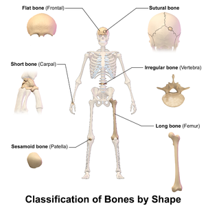
There are v types of bones in the man body: long, brusque, flat, irregular, and sesamoid.[29]
- Long bones are characterized by a shaft, the diaphysis, that is much longer than its width; and by an epiphysis, a rounded head at each end of the shaft. They are made up generally of compact bone, with bottom amounts of marrow, located within the medullary cavity, and areas of spongy, cancellous bone at the ends of the basic.[30] Most basic of the limbs, including those of the fingers and toes, are long bones. The exceptions are the eight carpal bones of the wrist, the vii articulating tarsal basic of the ankle and the sesamoid bone of the kneecap. Long bones such as the clavicle, that have a differently shaped shaft or ends are as well called modified long bones.
- Short bones are roughly cube-shaped, and have simply a thin layer of compact bone surrounding a spongy interior. The bones of the wrist and ankle are brusque bones.
- Flat bones are thin and by and large curved, with ii parallel layers of compact bone sandwiching a layer of spongy os. Well-nigh of the bones of the skull are flat bones, equally is the sternum.[31]
- Sesamoid bones are basic embedded in tendons. Since they deed to hold the tendon further away from the joint, the angle of the tendon is increased and thus the leverage of the muscle is increased. Examples of sesamoid bones are the patella and the pisiform.[32]
- Irregular bones practice non fit into the above categories. They consist of sparse layers of compact os surrounding a spongy interior. Equally implied by the name, their shapes are irregular and complicated. Oft this irregular shape is due to their many centers of ossification or because they contain bony sinuses. The bones of the spine, pelvis, and some bones of the skull are irregular bones. Examples include the ethmoid and sphenoid bones.[33]
Terminology [edit]
In the study of anatomy, anatomists utilise a number of anatomical terms to describe the advent, shape and office of bones. Other anatomical terms are likewise used to describe the location of bones. Similar other anatomical terms, many of these derive from Latin and Greek. Some anatomists nevertheless apply Latin to refer to bones. The term "osseous", and the prefix "osteo-", referring to things related to bone, are still used commonly today.
Some examples of terms used to depict basic include the term "foramen" to describe a hole through which something passes, and a "canal" or "meatus" to describe a tunnel-like structure. A protrusion from a bone tin exist called a number of terms, including a "condyle", "crest", "spine", "eminence", "tubercle" or "tuberosity", depending on the protrusion'south shape and location. In general, long bones are said to take a "head", "cervix", and "trunk".
When ii basic bring together together, they are said to "articulate". If the two bones take a fibrous connection and are relatively immobile, then the joint is chosen a "suture".
Development [edit]

Endochondral ossification
Light micrograph of a section through a juvenile human knee joint (rat) showing the cartilagineous growth plates
The formation of os is called ossification. During the fetal stage of development this occurs by 2 processes: intramembranous ossification and endochondral ossification.[34] Intramembranous ossification involves the formation of bone from connective tissue whereas endochondral ossification involves the germination of bone from cartilage.
Intramembranous ossification mainly occurs during formation of the apartment bones of the skull but too the mandible, maxilla, and clavicles; the bone is formed from connective tissue such equally mesenchyme tissue rather than from cartilage. The process includes: the development of the ossification centre, calcification, trabeculae formation and the development of the periosteum.[35]
Endochondral ossification occurs in long basic and about other bones in the body; it involves the development of bone from cartilage. This process includes the development of a cartilage model, its growth and development, development of the principal and secondary ossification centers, and the germination of articular cartilage and the epiphyseal plates.[36]
Endochondral ossification begins with points in the cartilage called "chief ossification centers." They more often than not appear during fetal development, though a few brusk bones begin their chief ossification afterward birth. They are responsible for the formation of the diaphyses of long bones, short bones and certain parts of irregular basic. Secondary ossification occurs later on birth and forms the epiphyses of long basic and the extremities of irregular and apartment bones. The diaphysis and both epiphyses of a long bone are separated by a growing zone of cartilage (the epiphyseal plate). At skeletal maturity (18 to 25 years of age), all of the cartilage is replaced by bone, fusing the diaphysis and both epiphyses together (epiphyseal closure).[37] In the upper limbs, only the diaphyses of the long bones and scapula are ossified. The epiphyses, carpal bones, coracoid process, medial border of the scapula, and acromion are notwithstanding cartilaginous.[38]
The post-obit steps are followed in the conversion of cartilage to bone:
- Zone of reserve cartilage. This region, farthest from the marrow cavity, consists of typical hyaline cartilage that equally yet shows no sign of transforming into bone.[39]
- Zone of cell proliferation. A little closer to the marrow crenel, chondrocytes multiply and arrange themselves into longitudinal columns of flattened lacunae.[39]
- Zone of prison cell hypertrophy. Next, the chondrocytes stop to split and brainstorm to hypertrophy (overstate), much like they do in the primary ossification center of the fetus. The walls of the matrix between lacunae become very thin.[39]
- Zone of calcification. Minerals are deposited in the matrix between the columns of lacunae and calcify the cartilage. These are not the permanent mineral deposits of bone, simply only a temporary support for the cartilage that would otherwise soon be weakened by the breakdown of the enlarged lacunae.[39]
- Zone of os degradation. Within each column, the walls between the lacunae break down and the chondrocytes die. This converts each cavalcade into a longitudinal aqueduct, which is immediately invaded by claret vessels and marrow from the marrow cavity. Osteoblasts line up along the walls of these channels and brainstorm depositing concentric lamellae of matrix, while osteoclasts dissolve the temporarily calcified cartilage.[39]
Functions [edit]
| Functions of os |
|---|
Mechanical
|
Constructed
|
Metabolic
|
Basic accept a multifariousness of functions:
Mechanical [edit]
Bones serve a diversity of mechanical functions. Together the bones in the torso grade the skeleton. They provide a frame to go along the body supported, and an attachment betoken for skeletal muscles, tendons, ligaments and joints, which function together to generate and transfer forces so that individual body parts or the whole body tin can be manipulated in 3-dimensional space (the interaction between bone and musculus is studied in biomechanics).
Bones protect internal organs, such as the skull protecting the brain or the ribs protecting the heart and lungs. Because of the way that bone is formed, bone has a loftier compressive strength of about 170 MPa (i,700 kgf/cm2),[8] poor tensile force of 104–121 MPa, and a very low shear stress force (51.6 MPa).[forty] [41] This means that bone resists pushing (compressional) stress well, resist pulling (tensional) stress less well, but only poorly resists shear stress (such equally due to torsional loads). While bone is essentially brittle, bone does accept a meaning degree of elasticity, contributed chiefly by collagen.
Mechanically, bones as well accept a special role in hearing. The ossicles are iii small bones in the middle ear which are involved in sound transduction.
Constructed [edit]
The cancellous part of bones comprise bone marrow. Bone marrow produces blood cells in a process called hematopoiesis.[42] Blood cells that are created in os marrow include ruby-red blood cells, platelets and white blood cells.[43] Progenitor cells such as the hematopoietic stem prison cell split in a process called mitosis to produce precursor cells. These include precursors which eventually requite rise to white claret cells, and erythroblasts which requite ascent to red blood cells.[44] Unlike cherry-red and white blood cells, created by mitosis, platelets are shed from very big cells chosen megakaryocytes.[45] This process of progressive differentiation occurs inside the bone marrow. Later the cells are matured, they enter the apportionment.[46] Every day, over ii.five billion cherry blood cells and platelets, and 50–100 billion granulocytes are produced in this way.[17]
Besides every bit creating cells, bone marrow is too one of the major sites where defective or aged blood-red blood cells are destroyed.[17]
Metabolic [edit]
- Mineral storage – bones act as reserves of minerals important for the body, most notably calcium and phosphorus.[47] [ commendation needed ] [48]
Adamant by the species, age, and the type of bone, bone cells make up to 15 per centum of the os. Growth gene storage—mineralized os matrix stores of import growth factors such as insulin-like growth factors, transforming growth factor, os morphogenetic proteins and others.[49]
- Fatty storage – marrow adipose tissue (MAT) acts as a storage reserve of fatty acids.[50]
- Acid-base balance – bone buffers the blood against excessive pH changes by absorbing or releasing alkaline salts.[51]
- Detoxification – bone tissues can also store heavy metals and other foreign elements, removing them from the blood and reducing their effects on other tissues. These tin can later be gradually released for excretion.[52]
- Endocrine organ – os controls phosphate metabolism past releasing fibroblast growth factor 23 (FGF-23), which acts on kidneys to reduce phosphate reabsorption. Bone cells also release a hormone chosen osteocalcin, which contributes to the regulation of blood sugar (glucose) and fat deposition. Osteocalcin increases both the insulin secretion and sensitivity, in addition to boosting the number of insulin-producing cells and reducing stores of fat.[53]
- Calcium balance – the process of bone resorption by the osteoclasts releases stored calcium into the systemic circulation and is an important procedure in regulating calcium balance. Equally bone formation actively fixes circulating calcium in its mineral course, removing it from the bloodstream, resorption actively unfixes it thereby increasing circulating calcium levels. These processes occur in tandem at site-specific locations.[54]
Remodeling [edit]
Bone is constantly being created and replaced in a process known as remodeling. This ongoing turnover of bone is a process of resorption followed past replacement of bone with petty change in shape. This is accomplished through osteoblasts and osteoclasts. Cells are stimulated by a variety of signals, and together referred to as a remodeling unit of measurement. Approximately 10% of the skeletal mass of an adult is remodelled each year.[55] The purpose of remodeling is to regulate calcium homeostasis, repair microdamaged bones from everyday stress, and to shape the skeleton during growth.[56] Repeated stress, such equally weight-bearing exercise or os healing, results in the bone thickening at the points of maximum stress (Wolff's law). It has been hypothesized that this is a result of bone's piezoelectric properties, which cause os to generate small electrical potentials under stress.[57]
The action of osteoblasts and osteoclasts are controlled by a number of chemical enzymes that either promote or inhibit the activity of the bone remodeling cells, controlling the charge per unit at which bone is made, destroyed, or changed in shape. The cells besides utilize paracrine signalling to control the activity of each other.[58] [59] For example, the rate at which osteoclasts resorb bone is inhibited by calcitonin and osteoprotegerin. Calcitonin is produced by parafollicular cells in the thyroid gland, and can bind to receptors on osteoclasts to directly inhibit osteoclast activity. Osteoprotegerin is secreted past osteoblasts and is able to bind RANK-50, inhibiting osteoclast stimulation.[60]
Osteoblasts can also be stimulated to increase bone mass through increased secretion of osteoid and by inhibiting the ability of osteoclasts to pause down osseous tissue.[ citation needed ] Increased secretion of osteoid is stimulated past the secretion of growth hormone past the pituitary, thyroid hormone and the sex hormones (estrogens and androgens). These hormones too promote increased secretion of osteoprotegerin.[lx] Osteoblasts can as well be induced to secrete a number of cytokines that promote reabsorption of bone by stimulating osteoclast activity and differentiation from progenitor cells. Vitamin D, parathyroid hormone and stimulation from osteocytes induce osteoblasts to increment secretion of RANK-ligand and interleukin 6, which cytokines then stimulate increased reabsorption of bone by osteoclasts. These aforementioned compounds also increment secretion of macrophage colony-stimulating factor by osteoblasts, which promotes the differentiation of progenitor cells into osteoclasts, and decrease secretion of osteoprotegerin.[ citation needed ]
Book [edit]
Bone volume is determined past the rates of os formation and bone resorption. Recent enquiry has suggested that certain growth factors may work to locally alter bone formation past increasing osteoblast activity. Numerous os-derived growth factors accept been isolated and classified via bone cultures. These factors include insulin-like growth factors I and Two, transforming growth gene-beta, fibroblast growth factor, platelet-derived growth factor, and os morphogenetic proteins.[61] Bear witness suggests that os cells produce growth factors for extracellular storage in the os matrix. The release of these growth factors from the bone matrix could cause the proliferation of osteoblast precursors. Essentially, bone growth factors may act equally potential determinants of local bone formation.[61] Research has suggested that cancellous os volume in postmenopausal osteoporosis may be determined past the relationship between the total os forming surface and the percent of surface resorption.[62]
Clinical significance [edit]
A number of diseases can affect bone, including arthritis, fractures, infections, osteoporosis and tumors. Weather condition relating to bone tin can be managed by a variety of doctors, including rheumatologists for joints, and orthopedic surgeons, who may carry surgery to fix cleaved bones. Other doctors, such as rehabilitation specialists may be involved in recovery, radiologists in interpreting the findings on imaging, and pathologists in investigating the cause of the illness, and family doctors may play a role in preventing complications of bone disease such equally osteoporosis.
When a doctor sees a patient, a history and exam volition be taken. Bones are and then oft imaged, called radiography. This might include ultrasound X-ray, CT scan, MRI scan and other imaging such as a Os browse, which may be used to investigate cancer.[63] Other tests such as a blood exam for autoimmune markers may exist taken, or a synovial fluid aspirate may exist taken.[63]
Fractures [edit]

In normal bone, fractures occur when at that place is pregnant force applied or repetitive trauma over a long time. Fractures tin also occur when a os is weakened, such as with osteoporosis, or when there is a structural problem, such as when the os remodels excessively (such as Paget'south disease) or is the site of the growth of cancer.[64] Common fractures include wrist fractures and hip fractures, associated with osteoporosis, vertebral fractures associated with high-free energy trauma and cancer, and fractures of long-basic. Not all fractures are painful.[64] When serious, depending on the fractures type and location, complications may include flail chest, compartment syndromes or fat embolism. Compound fractures involve the bone'southward penetration through the skin. Some complex fractures can exist treated past the employ of bone grafting procedures that replace missing bone portions.
Fractures and their underlying causes can exist investigated by X-rays, CT scans and MRIs.[64] Fractures are described by their location and shape, and several classification systems exist, depending on the location of the fracture. A common long bone fracture in children is a Salter–Harris fracture.[65] When fractures are managed, hurting relief is often given, and the fractured surface area is oftentimes immobilised. This is to promote os healing. In addition, surgical measures such as internal fixation may be used. Considering of the immobilisation, people with fractures are often advised to undergo rehabilitation.[64]
Tumors [edit]
There are several types of tumor that can affect os; examples of benign bone tumors include osteoma, osteoid osteoma, osteochondroma, osteoblastoma, enchondroma, giant-cell tumor of bone, and aneurysmal bone cyst.[66]
Cancer [edit]
Cancer can ascend in bone tissue, and basic are also a common site for other cancers to spread (metastasise) to.[67] Cancers that arise in os are called "chief" cancers, although such cancers are rare.[67] Metastases within bone are "secondary" cancers, with the near common beingness breast cancer, lung cancer, prostate cancer, thyroid cancer, and kidney cancer.[67] Secondary cancers that affect os can either destroy os (called a "lytic" cancer) or create bone (a "sclerotic" cancer). Cancers of the bone marrow within the bone can also affect os tissue, examples including leukemia and multiple myeloma. Bone may also be affected by cancers in other parts of the trunk. Cancers in other parts of the torso may release parathyroid hormone or parathyroid hormone-related peptide. This increases bone reabsorption, and tin lead to bone fractures.
Bone tissue that is destroyed or altered as a result of cancers is distorted, weakened, and more prone to fracture. This may atomic number 82 to compression of the spinal cord, destruction of the marrow resulting in bruising, bleeding and immunosuppression, and is 1 cause of bone pain. If the cancer is metastatic, and then there might be other symptoms depending on the site of the original cancer. Some bone cancers tin can also exist felt.
Cancers of the bone are managed according to their type, their phase, prognosis, and what symptoms they cause. Many primary cancers of bone are treated with radiotherapy. Cancers of bone marrow may be treated with chemotherapy, and other forms of targeted therapy such as immunotherapy may be used.[68] Palliative care, which focuses on maximising a person'south quality of life, may play a role in management, particularly if the likelihood of survival within five years is poor.
Other painful conditions [edit]
- Osteomyelitis is inflammation of the bone or bone marrow due to bacterial infection.[69]
- Osteomalacia is a painful softening of adult bone caused past severe vitamin D deficiency.[lxx]
- Osteogenesis imperfecta[71]
- Osteochondritis dissecans[72]
- Ankylosing spondylitis[73]
- Skeletal fluorosis is a os illness acquired by an excessive accumulation of fluoride in the bones. In advanced cases, skeletal fluorosis damages bones and joints and is painful.[74]
Osteoporosis [edit]
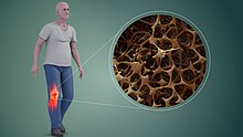
Reduced bone mineral density in Osteoporosis (R), increasing the likelihood of fractures
Osteoporosis is a disease of bone where there is reduced bone mineral density, increasing the likelihood of fractures.[75] Osteoporosis is divers in women by the World Health Organization as a bone mineral density of 2.5 standard deviations below peak bone mass, relative to the age and sex-matched boilerplate. This density is measured using dual free energy X-ray absorptiometry (DEXA), with the term "established osteoporosis" including the presence of a fragility fracture.[76] Osteoporosis is nearly common in women after menopause, when it is called "postmenopausal osteoporosis", only may develop in men and premenopausal women in the presence of particular hormonal disorders and other chronic diseases or every bit a event of smoking and medications, specifically glucocorticoids.[75] Osteoporosis usually has no symptoms until a fracture occurs.[75] For this reason, DEXA scans are often done in people with one or more than risk factors, who have developed osteoporosis and are at hazard of fracture.[75]
Osteoporosis treatment includes communication to stop smoking, decrease alcohol consumption, exercise regularly, and take a salubrious diet. Calcium and trace mineral supplements may likewise be advised, as may Vitamin D. When medication is used, it may include bisphosphonates, Strontium ranelate, and hormone replacement therapy.[77]
Osteopathic medicine [edit]
Osteopathic medicine is a school of medical idea originally developed based on the thought of the link between the musculoskeletal system and overall health, but now very similar to mainstream medicine. Equally of 2012[update], over 77,000 physicians in the United States are trained in osteopathic medical schools.[78]
Osteology [edit]

Human femurs and humerus from Roman catamenia, with prove of healed fractures
The study of bones and teeth is referred to every bit osteology. It is frequently used in anthropology, archeology and forensic science for a diverseness of tasks. This can include determining the nutritional, wellness, age or injury status of the private the bones were taken from. Preparing fleshed basic for these types of studies can involve the procedure of maceration.
Typically anthropologists and archeologists study os tools made by Human being sapiens and Homo neanderthalensis. Bones can serve a number of uses such as projectile points or creative pigments, and can also be made from external bones such as antlers.
Other animals [edit]


Leg and pelvic girdle bones of bird
Bird skeletons are very lightweight. Their basic are smaller and thinner, to aid flight. Among mammals, bats come closest to birds in terms of os density, suggesting that minor dumbo bones are a flight accommodation. Many bird basic accept little marrow due to their existence hollow.[79]
A bird'due south beak is primarily made of bone as projections of the mandibles which are covered in keratin.
Some basic, primarily formed separately in subcutaneous tissues, include headgears (such as bony core of horns, antlers, ossicones), osteoderm, and os penis/ os clitoris.[80] A deer's antlers are composed of bone which is an unusual example of bone being exterior the skin of the animal once the velvet is shed.[81]
The extinct predatory fish Dunkleosteus had sharp edges of hard exposed bone along its jaws.[82] [83]
The proportion of cortical bone that is 80% in the human skeleton may be much lower in other animals, peculiarly in marine mammals and marine turtles, or in various Mesozoic marine reptiles, such as ichthyosaurs,[84] amidst others.[85] This proportion tin can vary quickly in evolution; information technology often increases in early stages of returns to an aquatic lifestyle, as seen in early whales and pinnipeds, amid others. Information technology subsequently decreases in pelagic taxa, which typically learn spongy bone, but aquatic taxa that live in shallow water tin can retain very thick, pachyostotic,[86] osteosclerotic, or pachyosteosclerotic[87] bones, specially if they motility slowly, similar sea cows. In some cases, fifty-fifty marine taxa that had acquired spongy os can revert to thicker, compact bones if they go adjusted to live in shallow water, or in hypersaline (denser) water.[88] [89] [90]
Many animals, particularly herbivores, do osteophagy—the eating of bones. This is presumably carried out in order to replenish lacking phosphate.
Many os diseases that affect humans also touch on other vertebrates—an example of one disorder is skeletal fluorosis.
Gild and culture [edit]
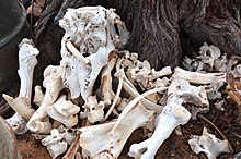
Bones from slaughtered animals accept a number of uses. In prehistoric times, they have been used for making bone tools.[91] They have farther been used in bone carving, already important in prehistoric art, and also in mod time every bit crafting materials for buttons, chaplet, handles, bobbins, calculation aids, head basics, dice, poker chips, choice-up sticks, arrows, scrimshaw, ornaments, etc.
Bone glue can be made by prolonged boiling of ground or croaky bones, followed past filtering and evaporation to thicken the resulting fluid. Historically in one case important, os mucilage and other fauna glues today have only a few specialized uses, such as in antiques restoration. Essentially the same process, with further refinement, thickening and drying, is used to brand gelatin.
Goop is made past simmering several ingredients for a long time, traditionally including basic.
Os char, a porous, blackness, granular cloth primarily used for filtration and likewise as a black pigment, is produced by charring mammal basic.
Oracle bone script was a writing system used in Ancient Red china based on inscriptions in bones. Its proper noun originates from oracle bones, which were mainly ox clavicle. The Ancient Chinese (mainly in the Shang dynasty), would write their questions on the oracle bone, and fire the bone, and where the os croaky would be the answer for the questions.
To point the bone at someone is considered bad luck in some cultures, such as Australian aborigines, such as by the Kurdaitcha.
The wishbones of fowl take been used for divination, and are still customarily used in a tradition to determine which 1 of 2 people pulling on either prong of the bone may make a wish.
Various cultures throughout history have adopted the custom of shaping an infant'due south head by the practice of artificial cranial deformation. A widely practised custom in Mainland china was that of pes bounden to limit the normal growth of the foot.
Additional images [edit]
-
Cells in bone marrow
-

Scanning electron microscope of bone at 100× magnification
-
Structure detail of an brute os
Run into also [edit]
- Artificial bone
- Bone health
- Distraction osteogenesis
- National Bone Health Campaign
- Skeleton
References [edit]
- ^ Lee, Cassandra. "The Bone Organ System: Course and Function". Scientific discipline Straight . Retrieved thirty January 2022.
- ^ de Buffrénil, Vivian; de Ricqlès, Armand J; Zylberberg, Louise; Padian, Kevin; Laurin, Michel; Quilhac, Alexandra (2021). Vertebrate skeletal histology and paleohistology (Firstiton ed.). Boca Raton, FL: CRC Press. pp. xii + 825. ISBN978-1351189576.
- ^ "How Many Bones Does a Baby Have and Why Do Adults Have Fewer?". Healthline. 26 June 2019. Retrieved 19 July 2021.
- ^ Steele, D. Gentry; Claud A. Bramblett (1988). The Anatomy and Biology of the Human Skeleton . Texas A&M Academy Press. p. 4. ISBN978-0-89096-300-5.
- ^ Mammal anatomy : an illustrated guide. New York: Marshall Cavendish. 2010. p. 129. ISBN9780761478829.
- ^ "ossein". The Gratuitous Lexicon.
- ^ Hall, John (2011). Textbook of Medical Physiology (twelfth ed.). Philadelphia: Elsevier. pp. 957–960. ISBN978-08089-2400-5.
- ^ a b Schmidt-Nielsen, Knut (1984). Scaling: Why Is Fauna Size So Important?. Cambridge: Cambridge Academy Press. p. half dozen. ISBN978-0-521-31987-four.
- ^ "Structure of Os". flexbooks.ck12.org. CK12-Foundation. Retrieved 28 May 2020.
- ^ a b c Deakin 2006, p. 192.
- ^ Gdyczynski, C.M.; Manbachi, A.; et al. (2014). "On estimating the directionality distribution in pedicle trabecular bone from micro-CT images". Physiological Measurement. 35 (12): 2415–2428. Bibcode:2014PhyM...35.2415G. doi:ten.1088/0967-3334/35/12/2415. PMID 25391037.
- ^ a b Deakin 2006, p. 195.
- ^ Hall, Susan J. (2007). Bones Biomechanics with OLC (5th ed., Revised. ed.). Burr Ridge: McGraw-Loma College Instruction. p. 88. ISBN978-0-07-126041-1.
- ^ Gomez, Santiago (February 2002). "Crisóstomo Martinez, 1638–1694: the discoverer of trabecular os". Endocrine. 17 (1): 3–4. doi:10.1385/ENDO:17:1:03. ISSN 1355-008X. PMID 12014701. S2CID 46340228.
- ^ Barnes-Svarney, Patricia L.; Svarney, Thomas E. (2016). The Handy Anatomy Answer Book : Includes Physiology. Detroit: Visible Ink Press. pp. 90–91. ISBN9781578595426.
- ^ a b c Deakin 2006, p. 189.
- ^ a b c Deakin 2006, p. 58.
- ^ Deakin 2006, pp. 189–190.
- ^ Washington. "The O' Cells." Bone Cells. University of Washington, n.d. Spider web. 3 Apr. 2013.
- ^ Sims, Natalie A.; Vrahnas, Christina (2014). "Regulation of cortical and trabecular os mass by advice between osteoblasts, osteocytes and osteoclasts". Archives of Biochemistry and Biophysics. 561: 22–28. doi:10.1016/j.abb.2014.05.015. PMID 24875146.
- ^ a b Deakin 2006, p. 190.
- ^ Enhancement of Hydroxyapatite Dissolution Journal of Materials Science & Engineering science,38, 148-158
- ^ a b c d e f Hall 2005, p. 981.
- ^ a b Currey, John D. (2002). "The Construction of Os Tissue", pp. 12–xiv in Basic: Construction and Mechanics. Princeton Academy Press. Princeton, NJ. ISBN 9781400849505
- ^ Salentijn, Fifty. Biology of Mineralized Tissues: Cartilage and Bone, Columbia University College of Dental Medicine post-graduate dental lecture series, 2007
- ^ Royce, Peter M.; Steinmann, Crush (xiv April 2003). Connective Tissue and Its Heritable Disorders: Molecular, Genetic, and Medical Aspects. John Wiley & Sons. ISBN978-0-471-46117-3.
- ^ Bertazzo, South.; Bertran, C. A. (2006). "Morphological and dimensional characteristics of os mineral crystals". Bioceramics. 309–311 (Pt. one, 2): three–10. doi:10.4028/world wide web.scientific.cyberspace/kem.309-311.3. S2CID 136883011.
- ^ Bertazzo, S.; Bertran, C.A.; Camilli, J.A. (2006). "Morphological Label of Femur and Parietal Bone Mineral of Rats at Unlike Ages". Key Engineering Materials. 309–311: xi–14. doi:10.4028/www.scientific.net/kem.309-311.11. S2CID 135813389.
- ^ "Types of bone". mananatomy.com. Retrieved half-dozen February 2016.
- ^ "DoITPoMS – TLP Library Construction of os and implant materials – Structure and composition of bone". www.doitpoms.ac.uk.
- ^ Bart Clarke (2008), "Normal Bone Anatomy and Physiology", Clinical Journal of the American Club of Nephrology, 3 (Suppl 3): S131–S139, doi:10.2215/CJN.04151206, PMC3152283, PMID 18988698
- ^ Adriana Jerez; Susana Mangione; Virginia Abdala (2010), "Occurrence and distribution of sesamoid basic in squamates: a comparative approach", Acta Zoologica, 91 (3): 295–305, doi:x.1111/j.1463-6395.2009.00408.x
- ^ Pratt, Rebecca. "Bone equally an Organ". AnatomyOne. Amirsys, Inc. Archived from the original on xxx October 2019. Retrieved 28 September 2012.
- ^ OpenStax, Anatomy & Physiology. OpenStax CNX. Feb 26, 2022 http://cnx.org/contents/14fb4ad7-39a1-4eee-ab6e-3ef2482e3e22@8.24
- ^ "Bone Growth and Evolution | Biological science for Majors II". courses.lumenlearning.com . Retrieved 28 May 2020.
- ^ Tortora, Gerard J.; Derrickson, Bryan H. (2018). Principles of Beefcake and Physiology. John Wiley & Sons. ISBN978-1-119-44445-9.
- ^ "six.4B: Postnatal Bone Growth". Medicine LibreTexts. 19 July 2018. Retrieved 28 May 2020.
- ^ Agur, Anne (2009). Grant'south Atlas of Beefcake. Philadelphia: Lippincott, Williams, and Wilkins. p. 598. ISBN978-0-7817-7055-2.
- ^ a b c d due east Saladin, Kenneth (2012). Beefcake and Physiology: The Unity of Form and Function. New York: McGraw-Hill. p. 217. ISBN978-0-07-337825-ane.
- ^ Vincent, Kevin. "Topic 3: Construction and Mechanical Properties of Bone". BENG 112A Biomechanics, Winter Quarter, 2013. Section of Bioengineering, University of California. Archived from the original on 28 May 2018. Retrieved 24 March 2015.
- ^ Turner, C.H.; Wang, T.; Burr, D.B. (2001). "Shear Force and Fatigue Properties of Man Cortical Bone Determined from Pure Shear Tests". Calcified Tissue International. 69 (6): 373–378. doi:ten.1007/s00223-001-1006-1. PMID 11800235. S2CID 30348345.
- ^ Fernández, KS; de Alarcón, PA (December 2013). "Development of the hematopoietic system and disorders of hematopoiesis that present during infancy and early childhood". Pediatric Clinics of North America. 60 (half dozen): 1273–1289. doi:10.1016/j.pcl.2013.08.002. PMID 24237971.
- ^ Deakin 2006, pp. 60–61.
- ^ Deakin 2006, p. 60.
- ^ Deakin 2006, p. 57.
- ^ Deakin 2006, p. 46.
- ^ Doyle, Máire E.; January de Beur, Suzanne M. (2008). "The Skeleton: Endocrine Regulator of Phosphate Homeostasis". Current Osteoporosis Reports. 6 (4): 134–141. doi:10.1007/s11914-008-0024-6. PMID 19032923. S2CID 23298442.
- ^ Walker, Kristin. "Bone". Encyclopedia Britannica . Retrieved 5 October 2017.
- ^ Pv, Hauschka; Tl, Chen; Ae, Mavrakos (1988). "Polypeptide Growth Factors in Bone Matrix". Ciba Foundation Symposium. Novartis Foundation Symposia. 136: 207–225. doi:x.1002/9780470513637.ch13. ISBN9780470513637. PMID 3068010. Retrieved 28 May 2020.
- ^ Styner, Maya; Pagnotti, Gabriel 1000; McGrath, Cody; Wu, Xin; Sen, Buer; Uzer, Gunes; Xie, Zhihui; Zong, Xiaopeng; Styner, Martin A (one May 2017). "Exercise Decreases Marrow Adipose Tissue through β-Oxidation in Obese Running Mice". Journal of Bone and Mineral Research. 32 (8): 1692–1702. doi:10.1002/jbmr.3159. ISSN 1523-4681. PMC5550355. PMID 28436105.
- ^ Fogelman, Ignac; Gnanasegaran, Gopinath; Wall, Hans van der (2013). Radionuclide and Hybrid Os Imaging. Springer. ISBN978-3-642-02400-9.
- ^ "Bone". flipper.diff.org . Retrieved 28 May 2020.
- ^ Lee, Na Kyung; et al. (10 August 2007). "Endocrine Regulation of Energy Metabolism by the Skeleton". Jail cell. 130 (3): 456–469. doi:10.1016/j.prison cell.2007.05.047. PMC2013746. PMID 17693256.
- ^ Foundation, CK-12. "Bones". www.ck12.org . Retrieved 29 May 2020.
- ^ Manolagas, SC (April 2000). "Birth and decease of bone cells: basic regulatory mechanisms and implications for the pathogenesis and treatment of osteoporosis". Endocrine Reviews. 21 (2): 115–137. doi:x.1210/edrv.21.2.0395. PMID 10782361.
- ^ Hadjidakis DJ, Androulakis 2 (31 January 2007). "Os remodeling". Register of the New York Academy of Sciences. 1092: 385–396. doi:10.1196/annals.1365.035. PMID 17308163. S2CID 39878618. Retrieved 18 May 2020.
- ^ ed, Russell T. Woodburne ..., consulting (1999). Anatomy, physiology, and metabolic disorders (5. print. ed.). Summit, Due north.J.: Novartis Pharmaceutical Corp. pp. 187–189. ISBN978-0-914168-88-i.
- ^ Fogelman, Ignac; Gnanasegaran, Gopinath; Wall, Hans van der (2013). Radionuclide and Hybrid Bone Imaging. Springer. ISBN978-iii-642-02400-ix.
- ^ "Introduction to cell signaling (commodity)". Khan University . Retrieved 24 Dec 2020.
- ^ a b Boulpaep, Emile L.; Boron, Walter F. (2005). Medical physiology: a cellular and molecular arroyo. Philadelphia: Saunders. pp. 1089–1091. ISBN978-1-4160-2328-ix.
- ^ a b Baylink, D. J. (1991). "Bone growth factors". Clinical Orthopaedics and Related Research. 263 (263): 30–48. doi:10.1097/00003086-199102000-00004. PMID 1993386.
- ^ Nordin, Be; Aaron, J; Speed, R; Crilly, RG (eight August 1981). "Bone formation and resorption as the determinants of trabecular bone volume in postmenopausal osteoporosis". Lancet. 2 (8241): 277–279. doi:ten.1016/S0140-6736(81)90526-two. PMID 6114324. S2CID 29646037.
- ^ a b Davidson 2010, pp. 1059–1062.
- ^ a b c d Davidson 2010, p. 1068.
- ^ Salter RB, Harris WR (1963). "Injuries Involving the Epiphyseal Plate". J Bone Joint Surg Am. 45 (3): 587–622. doi:10.2106/00004623-196345030-00019. Archived from the original on 2 December 2016. Retrieved two December 2016.
- ^ "Benign Bone Tumours". Cleveland Clinic. 2017. Retrieved 29 March 2017.
- ^ a b c Davidson 2010, p. 1125.
- ^ Davidson 2010, p. 1032.
- ^ "Osteomyelitis". The Lecturio Medical Concept Library . Retrieved 26 August 2021.
- ^ "Osteomalacia and Rickets". The Lecturio Medical Concept Library . Retrieved 26 August 2021.
- ^ "Osteogenesis Imperfecta". The Lecturio Medical Concept Library . Retrieved 26 August 2021.
- ^ "Osteochondritis Dissecans". The Lecturio Medical Concept Library . Retrieved 26 August 2021.
- ^ "Ankylosing Spondylitis". The Lecturio Medical Concept Library . Retrieved 26 Baronial 2021.
- ^ Whitford GM (1994). "Intake and Metabolism of Fluoride". Advances in Dental Research. 8 (1): 5–14. doi:10.1177/08959374940080011001. PMID 7993560. S2CID 21763028.
- ^ a b c d Davidson 2010, pp. 1116–1121.
- ^ WHO (1994). "Cess of fracture risk and its application to screening for postmenopausal osteoporosis. Study of a WHO Report Group". World Health Arrangement Technical Written report Series. 843: 1–129. PMID 7941614.
- ^ Davidson 2010, pp. 1116–1121
- ^ "2012 Osteopathic Medical Profession Report" (PDF). Osteopathic.org. American Osteopathic Organisation. Archived from the original (PDF) on sixteen June 2013. Retrieved 26 November 2014.
- ^ Dumont, Due east. R. (17 March 2010). "Os density and the lightweight skeletons of birds". Proceedings of the Purple Society B: Biological Sciences. 277 (1691): 2193–2198. doi:x.1098/rspb.2010.0117. PMC2880151. PMID 20236981.
- ^ Nasoori, A (2020). "Germination, structure, and function of extra‐skeletal basic in mammals". Biological Reviews. 95 (4): 986–1019. doi:ten.1111/brv.12597. PMID 32338826. S2CID 216556342.
- ^ Hans J. Rolf; Alfred Enderle (1999). "Hard fallow deer antler: a living bone till antler casting?". The Anatomical Record. 255 (one): 69–77. doi:x.1002/(SICI)1097-0185(19990501)255:1<69::Aid-AR8>iii.0.CO;2-R. PMID 10321994.
- ^ "Dunkleosteus". American Museum of Natural History.
- ^ "My, What a Large Rima oris You Accept | Cleveland Museum of Natural History".
- ^ de Buffrénil Five.; Mazin J.-M. (1990). "Bone histology of the ichthyosaurs: comparative data and functional interpretation". Paleobiology. 16 (four): 435–447. doi:x.1017/S0094837300010174. JSTOR 2400968.
- ^ Laurin, Chiliad.; Canoville, A.; Germain, D. (2011). "Bone microanatomy and lifestyle: a descriptive approach". Comptes Rendus Palevol. x (v–6): 381–402. doi:10.1016/j.crpv.2011.02.003.
- ^ Houssaye, Alexandra; De Buffrenil, Vivian; Rage, Jean-Claude; Bardet, Nathalie (12 September 2008). "An analysis of vertebral 'pachyostosis' in Carentonosaurus mineaui (Mosasauroidea, Squamata) from the Cenomanian (early Late Cretaceous) of France, with comments on its phylogenetic and functional significance". Journal of Vertebrate Paleontology. 28 (three): 685–691. doi:x.1671/0272-4634(2008)28[685:AAOVPI]2.0.CO;2. ISSN 0272-4634.
- ^ de Buffrénil, Vivian; Canoville, Aurore; D'Anastasio, Ruggero; Domning, Daryl P. (June 2010). "Evolution of Sirenian Pachyosteosclerosis, a Model-case for the Study of Bone Structure in Aquatic Tetrapods". Journal of Mammalian Evolution. 17 (2): 101–120. doi:x.1007/s10914-010-9130-1. S2CID 39169019.
- ^ Dewaele, Leonard; Lambert, Olivier; Laurin, Michel; De Kock, Tim; Louwye, Stephen; de Buffrénil, Vivian (December 2019). "Generalized Osteosclerotic Condition in the Skeleton of Nanophoca vitulinoides, a Dwarf Seal from the Miocene of Kingdom of belgium". Journal of Mammalian Development. 26 (4): 517–543. doi:10.1007/s10914-018-9438-9. S2CID 20885865.
- ^ Dewaele, Leonard; Gol'din, Pavel; Marx, Felix G.; Lambert, Olivier; Laurin, Michel; Obadă, Theodor; Buffrénil, Vivian de (10 January 2022). "Hypersalinity drives convergent os mass increases in Miocene marine mammals from the Paratethys". Current Biological science. 32 (ane): 248–255.e2. doi:10.1016/j.cub.2021.x.065. ISSN 0960-9822. PMID 34813730. S2CID 244485732.
- ^ Houssaye, Alexandra (10 January 2022). "Evolution: Back to heavy bones in salty seas". Current Biology. 32 (i): R42–R44. doi:10.1016/j.cub.2021.11.049. PMID 35015995. S2CID 245879886.
- ^ Laszlovszky, J¢zsef; Szab¢, P‚ter (i January 2003). People and Nature in Historical Perspective. Key European University Press. ISBN978-963-9241-86-2.
Footnotes [edit]
- Katja Hoehn; Marieb, Elaine Nicpon (2007). Homo Anatomy & Physiology (7th ed.). San Francisco: Benjamin Cummings. ISBN978-0-8053-5909-1.
- Bryan H. Derrickson; Tortora, Gerard J. (2005). Principles of beefcake and physiology. New York: Wiley. ISBN978-0-471-68934-8.
- Davidson, Stanley (2010). Colledge, Nicki R.; Walker, Brian R.; Ralston, Stuart H. (eds.). Davidson's principles and practise of medicine. Illustrated by Robert Britton (21st ed.). Edinburgh: Churchill Livingstone/Elsevier. ISBN978-0-7020-3085-vii.
- Deakin, Barbara Young; et al. (2006). Wheater's functional histology : a text and color atlas (5th ed.). [Edinburgh?]: Churchill Livingstone/Elsevier. ISBN978-0-443-068-508. – drawings by Philip J.
- Hall, Arthur C.; Guyton, John Due east. (2005). Textbook of medical physiology (11th ed.). Philadelphia: W.B. Saunders. ISBN978-0-7216-0240-0.
- Anthony, Due south. Fauci; Harrison, T.R.; et al. (2008). Harrison's principles of internal medicine (17th ed.). New York [etc.]: McGraw-Hill Medical. ISBN978-0-07-147692-8. – Anthony edits the current version; Harrison edited previous versions.
External links [edit]
| | Wikimedia Eatables has media related to Bones. |
| | Wikiquote has quotations related to: Bone |
- Educational resource materials (including animations) by the American Order for Bone and Mineral Research
- Review (including references) of piezoelectricity and bone remodelling
- A expert basic overview of bone biological science from the Science Creative Quarterly
- Usha Kini; B. Northward. Nandeesh (iii January 2013). "Ch two: Physiology of Bone Formation, Remodeling, and Metabolism" (PDF). In Ignac Fogelman; Gopinath Gnanasegaran; Hans van der Wall (eds.). Radionuclide and hybrid os imaging. Berlin: Springer. pp. 29–57. ISBN978-3-642-02399-6.
- Bone histology photomicrographs
Source: https://en.wikipedia.org/wiki/Bone
0 Response to "Bones & Born Again Diane Cluck"
Postar um comentário